

AGM: Using Breathing Circuits and Ventilators
The Anesthesia Gas Machine 2026
Michael P. Dosch PhD CRNA (retired), Darin Tharp CRNA MS
Nurse Anesthesia, University of Detroit Mercy
Revised Feb 2026. © Copyright Michael P. Dosch. Use for non-profit purposes only is granted.

9. Using Breathing Circuits and Ventilators
- Humidification
- Choosing the best fresh gas flow
- Low flows
- How to denitrogenate ("preoxygenate")
- Malignant hyperthermia implications for equipment.
- Ventilator and Breathing circuit problems and hazards
- Increased inspired carbon dioxide (troubleshooting and treatment)
Humidification
Dry gas supplied by the gas machine may cause clinically significant desiccation of mucus and an impaired mucociliary elevator. This may contribute to retention of secretions, blocking of conducting airways, atelectasis, bacterial colonization, and pneumonia.
- Absolute humidity is the maximum mass of water vapor which can be carried by a given volume of air ( expressed as mg/L). This quantity is strongly determined by temperature (warm air can carry much more moisture).
- Relative humidity (RH) is the amount present in a sample, as compared to the absolute humidity possible at the sample temperature (expressed as a percentage).
Some examples:
- 0 mg/L are supplied by the machine,
- 9 mg/L is found in normal room air at 20 degrees C and 50% relative humidity,
- 44 mg/L is found in tracheal air at the carina, at 37 degrees C and 100% relative humidity.
It is ideal to provide gases at body temperature and 100% RH to the patient’s airway. For cases lasting longer than 1 hour, humidification measures are often employed including:
- Use the circle with carbon dioxide absorbent granules and low flows. This can provide 100% RH at room temperature at the lowest flows (such as closed circuit with FGF of less than 1 L/min).
- Heat and moisture exchanger ("artificial nose")
The heat and moisture exchanger has large thermal capacity, hygroscopic, and (sometimes) bacterial filtration. It can do no more than return the patient’s exhaled water- it can’t add heat or moisture- and it is less efficient with longer cases or higher flows. But it’s easy to use, inexpensive, silent, won’t overheat or overhydrate the patient.
How is the "best" fresh gas flow (FGF) determined?
FGF determines not just F IO 2, but also the speed with which you can change the composition of gases in the breathing circuit.
- FGF of 4 L/min was common formerly- a legacy from days when a safety margin was needed for flowmeters & vaporizers which were much less accurate, much larger, and much leakier breathing circuits.
- At a FGF of 1-1.5 times V E (minute ventilation) (at least 5-8 L/min for an adult), there is no rebreathing of exhaled gas in the circle breathing system. At that high FGF, exhaled gases are rapidly diluted and exit via the scavenger before any can return to the patient. FGF should be this high during preoxygenation and induction (allows washin) and emergence (washout). In three breathing circuits (Aestiva, Fabius, ADU), time to 90% change in gas composition was not greatly faster at 6 or 8 L than at 4 L/m fresh gas flow. [1] So it's probably unnecessary to go beyond 8 L/min to speed preoxygenation, induction, or emergence.
- Low flows (0.5-2 L/min total FGF) should be used during maintenance to conserve tracheal heat and humidity, and economize on volatile agents.
- Key points
- You may close down to low flows after the airway is secured, particularly if you are employing End-tidal Control of expired oxygen and anesthetic agent. Remember, brain concentration lags behind the end-tidal agent concentration displayed on the monitor (both at induction, and during emergence).
- Don’t fear low flows with sevoflurane or desflurane provided you use absorbent that doesn't produce Compound A or carbon monoxide. The package insert advises against it. But in an excellent recent article, a trio of experts explains why low flows are safe with sevoflurane. [2]
Low flows
Low flows are used to decrease the usage, cost, and pollution of volatile anesthetics. A 50% reduction in FGF translates to a 50% savings on gaseous anesthetic agent, without placing the patient at risk or lessening the quality of their care (savings are offset by quicker consumption of absorbent). Tracheal heat and humidity, and patient core body temperature are preserved better than at higher flows. Low flows are ecologically sound, lessening the release of volatile agents and nitrous oxide, with their global warming potential. [3]
The composition of gases in the breathing circuit may change as lower flows are employed, since a greater fraction of the gas inspired by the patient will be rebreathed.
- Oxygen: inspired oxygen may decline to less than the amount set on the flowmeter, especially as delivered oxygen approaches the metabolic requirement for oxygen (250-300 mL/min in an adult). This can readily and rapidly occur with low FGF and low set F IO 2. Inspired oxygen declines because of uptake, and dilution of oxygen distal to the common gas outlet by leaks, exhaled nitrogen, carbon dioxide, and water vapor. [4], [5], [6]
- Agent: inspired agent may be much less than that dialed on the vaporizer when low flows are employed, also due to leaks, uptake and dilution.
- These problems are addressed in new designs.
- The Maquet Flow-i workstation utilizes an active hypoxic guard, which overrules settings when F IO 2 declines towards a hypoxic breathing mixture. The Flow-i monitors inspired (not merely delivered oxygen). [7]
- The Aisys CS2 uses Endtidal Control. The user sets targets for minimum flow (FGF), expired oxygen, and expired anesthetic agent. The Aisys manipulates FGF and delivered agent concentrations to quickly and effectively achieve and maintain these targets. [8], [9] Maquet has a similar system, Automatic Gas Control with Active Hypoxic Guard. [10] Mindray A9 has a similar automatically controlled anesthesia (ACA) module. [11]
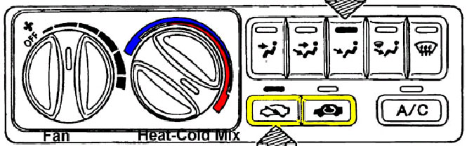
Car heater controls- fan and heat mix. Click on the link to see the larger version.
Large discrepancies between dialed and inspired agent concentration can be unsettling, raising apprehensions about vaporizer or breathing circuit malfunction. An analogy may help to clarify why this is an expected result of low flows.
Imagine you are entering an automobile in the winter. You turn the heater on at maximum heat level and fan speed. After the car is warmed to a comfortable temperature, you can't turn the heater entirely off. Heat must still be supplied, since it is always escaping (the car is not airtight). To keep the car at the desired temperature, you may either a) flow a moderate to high fan speed, but decrease the heat mix to nearly room temperature air, or b) you may leave the heat mix level high, and slowly blow in a small amount of very hot air. It makes no difference- in either case the car stays at the desired temperature.
Similarly, we begin cases with higher flows. Since there is little rebreathing at 4 L/min FGF and above, the dialed and inspired agent concentration are very similar. We induce with overpressure until the patient is saturated (reflected in an end-tidal agent concentration which closely approaches inspired). Then we may either a) leave the flows high with a moderate agent concentration near MAC, or b) turn the flows to low flow. But if we use low flows, we must still provide the same number of molecules of agent in order to replace that lost due to dilution, leaks, and uptake to fat and muscle. So with low FGF, we must turn the vaporizer dial beyond what we might have at higher flows.
Advantages of low flows
- Economy
- Decreased operating room & environmental pollution
- Estimation of agent uptake and oxygen consumption
- At some point below 1 L/m, low flows become closed circuit: the APL valve is closed and only enough gases and agent are supplied to keep the bellows or bag volume constant. One can then infer uptake from changes in volume and composition of the gases in the breathing circuit.
- Buffering of changes in inspired concentration
- Conservation of heat and humidity
Disadvantages of low flows
- More attention required
- If you are not using End-tidal Control, it is difficult to quickly alter inspired concentrations; if you must lighten or deepen the agent level quickly, switch for a moment to higher flows or use strong overpressure to change depth quickly.
- Danger of hypercarbia, expense; absorbent granules are used at a faster rate with low flows because of the higher degree of rebreathing
- Greater knowledge required (only if closed circuit employed)
- Accumulation of undesired gases in the circuit (only if closed circuit employed)
- Carbon monoxide, acetone, methane, hydrogen, ethanol, anesthetic agent metabolites, argon, nitrogen
Contraindications for low flows- Absolute and relative
There are two excellent comprehensive reviews of low flow anesthesia noted below. [12], [13]
- Absolute
- High VO 2 needed (e.g. Malignant Hyperthermia)
- Toxic gases to washout (e.g. smoke inhalation)
- Necessary equipment broken/missing (e.g. Oxygen analyzer, agent analyzer, absorbent)
- Relative
- Case duration < 15 minutes
- Leaks to be expected
- older gas machine (less leak-proof), face mask anesthesia or LMA, uncuffed endotracheal tubes, during rigid bronchoscopy (huge leaks)
How to denitrogenate ("preoxygenate")
You can "preoxygenate" with a nasal cannula or any other oxygen source. We need to do more- denitrogenate (cleanse the functional residual capacity of nitrogen)- to help our patients tolerate a potential period of apnea (if we have difficulties with intubation).
- Fresh gas flow 6-8 L/min
- APL valve open fully
- Tight mask fit- the most significant factor. It cannot be compensated for by increasing time of preoxygenation, because the patient will not be breathing 100% oxygen with a loose fit. [14]
- Every time you place a mask on a patient's face, look back at the breathing bag (to ensure it is fluctuating with respirations) and the oxygen flowmeter (to ensure it is on).
- Pay attention to complaints that it "smells funny"- you may have left a vaporizer on.
- Thus you can avoid the threats to patient safety inherent in an apneic patient (pretreatment with non-depolarizing muscle relaxants, relative overdose of narcotics), or one to whom oxygen is not being provided, or one exposed to anesthetic vapor before this is intended.
Malignant hyperthermia: Implications for equipment
Clinical presentation The cause of the symptoms (tachycardia, tachypnea, and elevated end-tidal CO 2) seen in malignant hyperthermia (MH) must be distinguished from ventilator or unidirectional valve malfunctions (producing respiratory acidosis), hyperthyroidism, cocaine intoxication, pheochromocytoma, and sepsis.
Triggers Succinylcholine and all inhaled agents are the only anesthetic agents that will trigger MH.
- Safe anesthetics: Barbiturates, propofol, etomidate, ketamine, opioids, local anesthetics, catecholamines, nitrous oxide, and all non-depolarizing muscle relaxants are presently considered safe. [15]
Treatment of acute episodes in OR High fresh gas flow (10 L/min), notify surgeon, hyperventilation, use activated charcoal filters on inspiratory and expiratory limbs of breathing circuit, stop inhaled agents and remove vaporizers, stop succinylcholine, and as time permits change soda lime granules & breathing circuit. The mainstay of treatment is dantrolene 2.5 mg/kg (up to 10 mg/kg). Cooling by any and all means, NaHCO 3, treatment of hyperkalemia, and other measures are also important. MHAUS (Malignant Hyperthermia Association of the US) publishes lists of recommendations for management of acute crisis at MHAUS.org.
Management of known susceptible patients- MHAUS recommends four alternatives to prepare the gas machine:
- Flush and prepare workstation according to manufacturer’s recommendations or published studies; this may take 10 to >90 minutes.
- Most studies also physically disconnect vaporizers from the workstation;
- use a new, disposable breathing circuit;
- and replace the carbon dioxide absorbent.
- During the case, FGF should be 10 L/miniters per minute to avoid “rebound phenomenon” (increased release of residual volatile anesthetic agent when fresh gas flow is reduced after a set period of flushing).
- OR Use commercially available activated charcoal filters following a 90 second flush with high fresh gas flows. These filters have been shown to remove trace levels of volatile anesthetic agents, and in one in vitro study to be effective for 12 hours.
- OR If available, use a dedicated “vapor free” machine for MH-susceptible patients. The machine must be regularly maintained and safety-checked.
- OR If appropriate to the institution, use an ICU ventilator that has never been exposed to volatile anesthetic agents.
- Note that guidelines for flushing (#1 above) do NOT apply to all machines. One study showed that a modern machine (Fabius GS) required 105 minutes of flushing before it was agent-free. [16]
Ventilator and Breathing Circuit: Problems and Hazards
Disconnection
Most common site is Y piece. The most common preventable equipment-related cause of mishaps. Direct your vigilance here by:
- use a precordial stethoscope
- if you turn the vent off temporarily (e.g. for an Xray), keep your finger on the switch
- use apnea alarms and don’t silence them.
- The biggest problem with ventilators is failure to initiate ventilation, or resume it after it is paused. Be extremely careful just after initiating ventilation- or whenever ventilation is interrupted: observe and listen to the chest for a few breathing cycles. Never take for granted that flipping the switches will cause ventilation to occur, or that you will always remember to turn the ventilator back on after an Xray.
Monitors for disconnection
- Precordial monitor (important because its "alarms" can't be inactivated). [17], [18], [19]
- Capnography
- Other monitors for disconnection
- Ascending bellows
- Observe chest excursion and epigastrium
- Airway pressure monitors
- Exhaled volume monitors
- Flow-time waveform
Occlusion/obstruction of breathing circuit
Beside inability to ventilate, obstruction may also lead to barotrauma. Obstruction may be related to:
- Foreign objects or manufacturing flaws that block all flow in the breathing circuit. If no flow test is done in the morning checklist and between patients, failure to ventilate may be misdiagnosed as bronchospasm on induction. The results have been, and continue to be, fatal. [20]
- Tracheal tube (kinked, biting down, plugged, or cuff balloon herniation). "All that wheezes is not bronchospasm".
- Incorrect insertion of flow-direction-sensitive components (older PEEP valves which are added on between the absorber head and corrugated breathing hoses)
- Excess inflow to breathing circuit (flushing during ventilator inspiratory cycle)
- Bellows leaks
- Ventilator relief valve (spill valve) malfunction
- APL valve too tight during mask ventilation or not fully open during preoxygenation.
Misconnection
Much less of a problem since breathing circuit and scavenger tubing sizes have been standardized. However, breathing systems are reconfigured for preventive maintenance and other reasons. One such incident resulted in apnea, inability to ventilate, and cardiac arrest (asystole) in 2007. [21]
Failure of emergency oxygen supply
May be due to failure to check cylinder contents, or driving a ventilator with cylinders when the pipeline is unavailable. This leads to their rapid depletion, perhaps in as little as an hour, since you need approximately a V T of driving gas per breath, substantially more if airway resistance (RAW) is increased. [22]
Infection
Clean the bellows after any patient with diseases which may be spread through airborne droplets, or don’t use the mechanical ventilator, or use bacterial filters, or use disposable soda lime assembly, or use a Bain.
Mechanical ventilator failure
Protocol for mechanical ventilator failure
- If the ventilator fails or continuing high pressure occurs, manually ventilate with the circle system. (Why? Allows you to continue using vaporizers.)
- If #1 is not possible, then bag with oxygen (if a portable cylinder is available) or room air (if not).
- If #2 is not possible, then try to pass suction catheter through the tracheal tube. ("When in doubt, pull it out" is not the best advice in all circumstances.)
- If #3 is not possible, then visualize the hypopharynx and cords with the tube in place, or possibly reintubate.
Increased inspired carbon dioxide
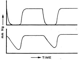
Inspired unidirectional valve problem- bottom capnogram. Click on the link to see the larger version.
The causes of increased inspired carbon dioxide are almost exclusively either malfunctioning unidirectional valves, or exhausted absorbent.
Increased inspired carbon dioxide has other potential causes, but these are rare
- inadvertent administration of carbon dioxide
- low fresh gas flow in a Mapleson system
- improper assembly of Bain system
- excessive dead space [for example rebreathing under drapes]
- leak in inspiratory limb of circle
- capnograph artifact [water in sampling cell, or sampling rate too low]
Many treatment approaches waste precious time: increasing minute ventilation; seeking signs of malignant hyperthermia; checking for leaks in the circuit; obtaining arterial blood gases; performing bronchoscopy for mucous plugs; inserting central lines; recalibrating or replacing ventilator, capnograph, or the entire gas machine.
Malfunctioning unidirectional valves can cause serious problems.
- If the inspiratory valve is incompetent, the patient exhales into both limbs. The capnogram may show a slanted downstroke inspiratory phase (as the patient inhales carbon dioxide-containing gas from the inspiratory limb) and increased end-tidal carbon dioxide (as in the bottom capnogram in the figure above).
- If the expiratory valve is incompetent, increased inhaled and exhaled carbon dioxide levels may appear with a normal appearing capnogram.
- The cardinal sign in either is an elevated baseline- a non-zero inspired CO 2.
- Failure of granules or valves has been defined as inspired CO 2 of 5 mm Hg. [24]
- Both situations result in respiratory acidosis unresponsive to increased ventilation.
- If the valves stick closed, all gas flow within the circle system ceases, and one cannot ventilate the patient.
- If either valve is stuck or obstructed, it causes immediate inability to ventilate (mechanically or manually). This must be differentiated from severe bronchospasm or endotracheal tube kinked or obstructed. Obstructed breathing circuits are checked as part of the morning checklist, and between patients using a flow test. See Checklist in a subsequent section of this site.
Differential diagnosis: Machine malfunction? Altered patient physiology?
Increased carbon dioxide production will not result in increased inspired carbon dioxide. The capacity of the soda lime granules is sufficient to cleanse each breath entirely, even if carbon dioxide production is increased. Further, respiratory acidosis will not cause visibly dark blood, or desaturation on the pulse oximeter.
Diagnosis and treatment
(Absent the rare causes mentioned above) if the granules are not exhausted, and the inspiratory and expiratory unidirectional valves are forcing all exhaled gas through the granules, there can be no increase in inspired carbon dioxide. So, if it is detected:
- First, increase fresh gas flow (FGF) to much greater than minute ventilation.
- A fresh gas flow of 8-10 L/min creates a semi-open system, with essentially no rebreathing, since the amount of fresh gas is sufficient to dilute any exhaled carbon dioxide to very low levels (and send it to the scavenging system).
- If the granules are exhausted, inspired CO 2 will return to normal when FGF is increased. Either leave FGF high and change granules at end of case, or change the granules as soon as practical and safe during the case, then return to low flow.
- If the increased fresh gas flow didn't decrease inspired CO 2, an incompetent valve is likely. The entire workstation should be replaced while bagging the patient, and assuring that anesthesia depth continues with TIVA. Modern breathing circuits do not allow correcting this fault while a patient is attached to the breathing circuit.
[1] Dosch MP, Loeb RG, Brainerd TL, Stallwood JF, Lechner S. Time to a 90% change in gas concentration: a comparison of three semi-closed anesthesia breathing systems. Anesth Analg. 2009 Apr;108(4):1193-7. doi: 10.1213/ane.0b013e3181949afd.
[2] Kennedy RR, Hendrickx JF, Feldman JM. There are no dragons: Low-flow anaesthesia with sevoflurane is safe. Anaesth Intensive Care. 2019 May;47(3):223-225. doi: 10.1177/0310057X19843304.
[3] ASA Committee on Equipment and Facilities (2023). Statement on the Use of Low Gas Flows for Sevoflurane. https://www.asahq.org/standards-and-practice-parameters/statement-on-the-use-of-low-gas-flows-for-sevoflurane, Accessed Feb 2, 2026
[4] Varughese S, Ahmed R. Environmental and Occupational Considerations of Anesthesia: A Narrative Review and Update. Anesth Analg. 2021 Oct 1;133(4):826-835. doi: 10.1213/ANE.0000000000005504.
[5] De Cooman S, Schollaert C, Hendrickx JF, Peyton PJ, Van Zundert T, De Wolf AM. Hypoxic guard systems do not prevent rapid hypoxic inspired mixture formation. J Clin Monit Comput. 2015 Aug;29(4):491-7. doi: 10.1007/s10877-014-9626-y. (especially video content)
[6] Eriksson S, Sixten B. More on the danger of inhaling air at reduced fresh gas flows: a self-experiment. J Clin Monit Comput. 2016 Apr;30(2):251-2. doi: 10.1007/s10877-015-9710-y.
[7] Ghijselings IE, De Cooman S, Carette R, Peyton PJ, De Wolf AM, Hendrickx JF. Performance of an active inspired hypoxic guard. J Clin Monit Comput. 2016 Feb;30(1):63-8. doi: 10.1007/s10877-015-9684-9.
[8] Wetz AJ, Mueller MM, Walliser K, Foest C, Wand S, Brandes IF, Waeschle RM, Bauer M. End-tidal control vs. manually controlled minimal-flow anesthesia: a prospective comparative trial. Acta Anaesthesiol Scand. 2017 Nov;61(10):1262-1269. doi: 10.1111/aas.12961.
[9] Singaravelu S, Barclay P. Automated control of end-tidal inhalation anaesthetic concentration using the GE Aisys Carestation™. Br J Anaesth. 2013 Apr;110(4):561-6. doi: 10.1093/bja/aes464.
[10] Getinge 2025. https://www.getinge.com/int/products-and-solutions/operating-room/anesthesia/AGC/ Accessed Feb 2, 2026
[11] Serefoglu R, Kocayigit H, Palabiyik O, Tuna AT. Comparison of automated and manual control methods in minimal flow anesthesia. J Clin Monit Comput. 2024 Oct;38(5):1117-1123. doi: 10.1007/s10877-024-01163-0.
[12] Hönemann C, Hagemann O, Doll D. Inhalational anaesthesia with low fresh gas flow. Indian J Anaesth. 2013 Jul;57(4):345-50. doi: 10.4103/0019-5049.118569.
[13] Honemann C, Mierke B. Low-flow, minimal-flow and metabolic-flow anaesthesia: Clinical techniques for use with rebreathing systems. 2015:Dräger (see page 86 for discussion of contraindications) at https://www.draeger.com/Content/Documents/Content/low-minimal-flow-anaesthesie-bk-9067990-en-global.pdf Accessed Feb 2, 2026
[14] Benumof JL. Preoxygenation: best method for both efficacy and efficiency. Anesthesiology. 1999 Sep;91(3):603-5. doi: 10.1097/00000542-199909000-00006.
[15] Malignant Hyperthermia Association of the US. Safe and Unsafe Anesthetics. https://www.mhaus.org/healthcare-professionals/be-prepared/safe-and-unsafe-anesthetics/ Accessed Feb , 2026
[16] Kim TW, Nemergut ME. Preparation of modern anesthesia workstations for malignant hyperthermia-susceptible patients: a review of past and present practice. Anesthesiology. 2011 Jan;114(1):205-12. doi: 10.1097/ALN.0b013e3181ee2cb7.
[17] Boriosi JP, Hollman GA. Making a case for use of the pretracheal stethoscope in pediatric procedural sedation. Paediatr Anaesth. 2016 Mar;26(3):249-55. doi: 10.1111/pan.12849.
[18] Fisher QA. Can capnography substitute for auscultation in sedation cases? Anesth Analg. 2005 May;100(5):1546. doi: 10.1213/01.ANE.0000151470.50760.4A.
[19] Lofsky AS. Turn Your Alarms On! APSF Newsletter 2004. https://www.apsf.org/article/turn-your-alarms-on/ Accessed Feb 2, 2026
[20] Mehta SP, Eisenkraft JB, Posner KL, Domino KB. Patient injuries from anesthesia gas delivery equipment: a closed claims update. Anesthesiology. 2013 Oct;119(4):788-95. doi: 10.1097/ALN.0b013e3182a10b5e.
[21] US FDA. Adverse event report MW5003097, Manufacturer and User Facility Device Experience [MAUDE] database, 2007
[22] Taenzer AH, Kovatsis PG, Raessler KL. E-cylinder-powered mechanical ventilation may adversely impact anesthetic management and efficiency. Anesth Analg. 2002 Jul;95(1):148-50, table of contents. doi: 10.1097/00000539-200207000-00026.
[23] Apfelbaum JL, Hagberg CA, Connis RT, Abdelmalak BB, Agarkar M, Dutton RP, Fiadjoe JE, Greif R, Klock PA, Mercier D, Myatra SN, O'Sullivan EP, Rosenblatt WH, Sorbello M, Tung A. 2022 American Society of Anesthesiologists Practice Guidelines for Management of the Difficult Airway. Anesthesiology. 2022 Jan 1;136(1):31-81. doi: 10.1097/ALN.0000000000004002.
[24] Feldman JM, Hendrickx J, Kennedy RR. Carbon Dioxide Absorption During Inhalation Anesthesia: A Modern Practice. Anesth Analg. 2021 Apr 1;132(4):993-1002. doi: 10.1213/ANE.0000000000005137.